×
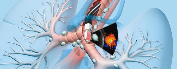
Endobronchial Ultrasound (EBUS) is an advanced, minimally invasive diagnostic procedure that uses a specialized bronchoscope fitted with an ultrasound probe to visualize the lungs and nearby lymph nodes in real time. This technology allows doctors to accurately locate abnormal areas and take precise tissue samples (biopsies) without any surgical cuts. EBUS plays a crucial role in diagnosing lung cancer, staging cancer spread, identifying infections like tuberculosis, and evaluating conditions such as sarcoidosis or unexplained lymph node enlargement. With its high accuracy, low risk, and quick recovery, EBUS has become one of the most reliable and comfortable techniques for evaluating chest diseases.